undefined
Similar Products


Description
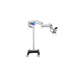
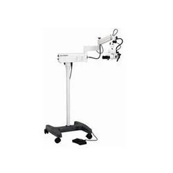
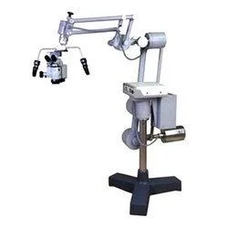
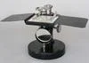
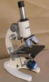
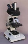
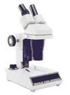
Our Premium Neuro Microscopes are precision optical instruments specifically designed for the detailed observation required in neurological studies and diagnostic procedures. They ...
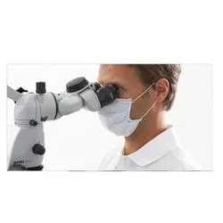
Our Premium Neuro Microscopes are precision optical instruments specifically designed for the detailed observation required in neurological studies and diagnostic procedures. They integrate high-grade optical components with a mechanically stable platform to deliver consistently clear and accurate images of neural tissues, cells, and pathological specimens. The system is built to perform reliably in environments where precision is paramount, featuring optimized illumination pathways and multiple contrast-enhancing options to reveal intricate structural details that are critical for both research conclusions and clinical assessments.

These microscopes serve as fundamental tools across sectors where understanding neural morphology and pathology is essential. In hospital laboratories, they are used by pathologists and technicians for examining tissue biopsies, aiding in the diagnosis of neurological disorders. University and private research institutes employ them for foundational and applied neuroscience, studying everything from basic neural circuits to disease mechanisms. The pharmaceutical and biotechnology industries rely on their precision for drug discovery workflows, toxicology studies, and quality control processes involving neural models or tissues.

The value proposition centers on delivering dependable performance that supports operational efficiency and confidence in results. Each microscope is subjected to stringent quality checks, aligning with the expectations for professional-grade laboratory equipment. This focus on reliability translates to reduced downtime and consistent data quality over the instrument's lifespan. The design prioritizes user ergonomics to minimize strain during long observation periods, thereby supporting productivity. The optical and mechanical quality ensures that investments are protected, providing a capable and lasting tool for critical observational tasks.

Key Features:
- Superior optical system with high-resolution objectives for detailed neural and cellular imaging.
- Robust and stable mechanical construction designed for consistent performance in laboratory settings.
- Versatile illumination system compatible with various contrast techniques like brightfield and phase contrast.
- Ergonomic design with adjustable components to reduce user fatigue during prolonged use.
- Modular design allowing integration with digital cameras and other imaging accessories for documentation.

Benefits:
- Achieve clear, detailed visualization of fine neural structures and pathological features for accurate analysis.
- Experience reliable operation with minimal maintenance, ensuring consistent availability for critical tasks.
- Adapt the instrument to various observation techniques and workflows without needing multiple specialized systems.
- Enhance operator comfort and efficiency, leading to more productive observation sessions and reduced error risk.
- Extend functionality by easily connecting to digital imaging systems for documentation, sharing, and quantitative analysis.
Specifications
| Additional Information | |
|---|---|
| Country of Origin | India |
| Customisable | No |
Application
These neuro microscopes are engineered for demanding neurological observation, providing exceptional clarity for studying neural tissue, cellular structures, and pathological samples. Their robust design ensures stable, vibration-free operation essential for capturing minute details during extended research sessions or critical diagnostic evaluations.

Medical and research institutions utilize these instruments for a wide range of observation protocols, from basic histology to advanced fluorescent imaging. The optical system supports various contrast methods and illumination techniques, making it adaptable for different specimen types and staining procedures without compromising image integrity or user comfort.
- Observing neural tissue architecture and cellular connectivity in academic neuroscience research.
- Analyzing biopsy samples and pathological specimens for accurate neurological disease diagnosis in clinical laboratories.
- Supporting intraoperative tissue examination and guidance during neurosurgical procedures.
- Conducting detailed cellular and molecular studies within pharmaceutical development and neuropharmacology research.
- Facilitating advanced training and education in medical schools and specialized neurological training programs.
Trade Details
| Available Stock | In stock |
| Sample Availability | No |
Payment Terms
| Payment Terms |
|
Company Profile
ABLE Healthcare Systems Private Limited, Punjab
 Punjab, India
Health & Personal Care•Trading CompanyRetailer
Factory Details
Factory SizeBelow 1000 sqm
Annual Production Capacity0
Premium Neuro Microscopes For Neurological Research And Diagnostics
Premium Neuro Microscopes deliver precise neurological imaging for research and diagnostics. Featuring superior optics and reliable construction, these instruments provide accurate high-performance observation trusted by laboratories and medical facilities.
Min. Order Quantity: 1 units
Shipping
Shipping fee and delivery date to be negotiated. Contact supplier now for more details.
ABLE Healthcare Systems Private Limited Punjab, India
Health & Personal CareTrading Company